We were tasked with creating an animation to show the benefits of the Paccocath technology – this meant developing different scenes (vessel views versus a close up cellular level) as well as static images to visualize the drug matrix.
Paccocath Short Animation
This is the short version of the animation, highlighting the benefits of the Paccocath technology. The full animation is built in sections, the first being a segment on standard balloon angioplasty and potential issues, and then other drug coated balloons and how they try to deal with those problems (this section is not shown in this short version). Finally we are introduced to the Paccocath technology and it’s unique approach.
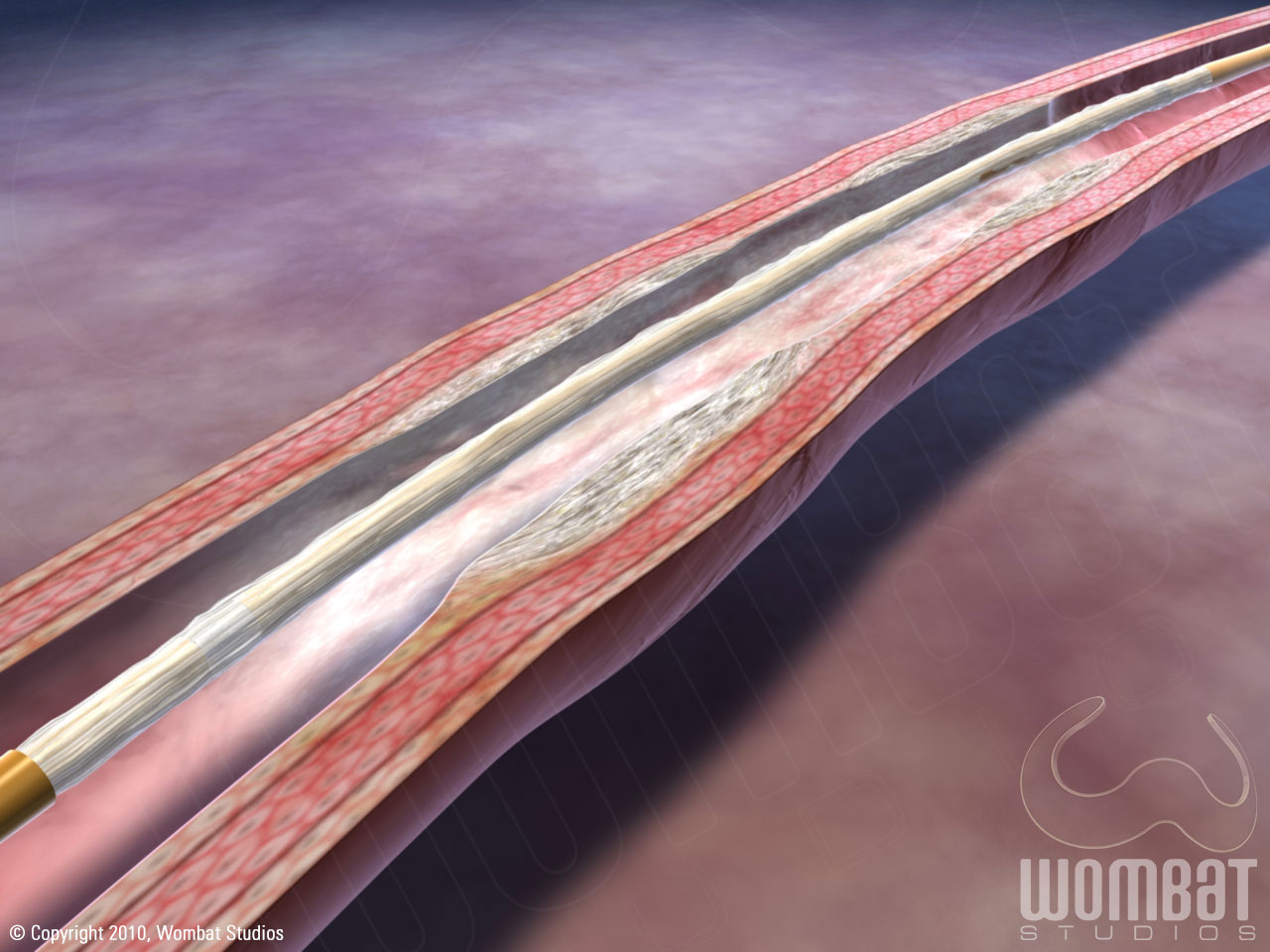
Paccocath: Prior to Balloon Inflation
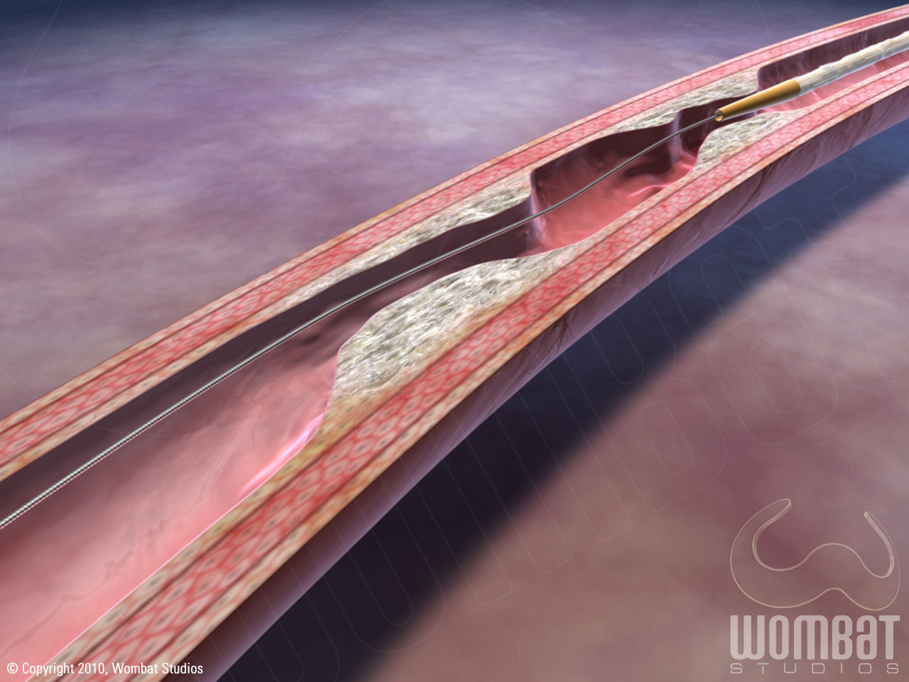
These are stills from the animation, what I call the “far away view” though it’s a very small catheter within a small blood vessel. This shows a stenotic calcified blood vessel with significant occlusion, and the Paccocath catheter being introduced to the lesion.
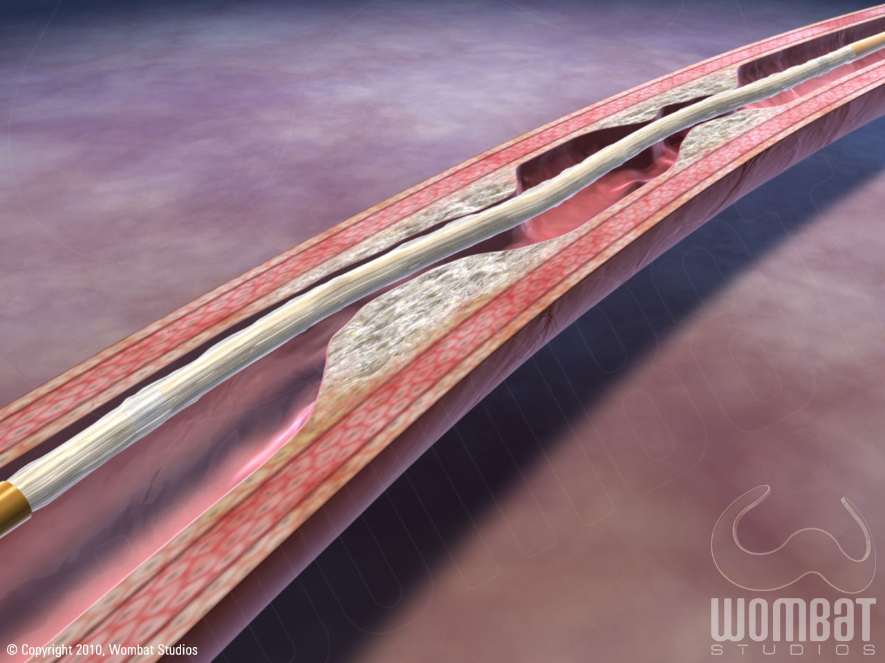
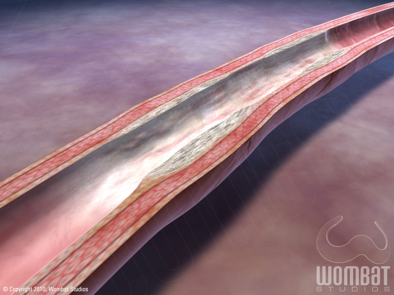
Paccocath: Inflated Balloon
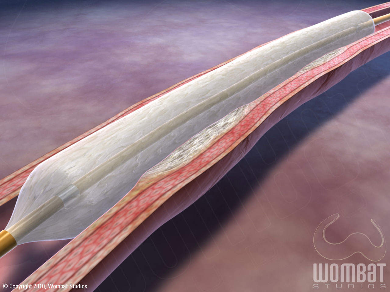
Another “far away view”; this is the Paccocath catheter balloon being inflated, expanding the vessel lumen (and the stenotic mass distending the vessel wall). The balloon remains expanded and pressed against the lesion, giving the drug coating time to stick and absorb into the lumen wall.
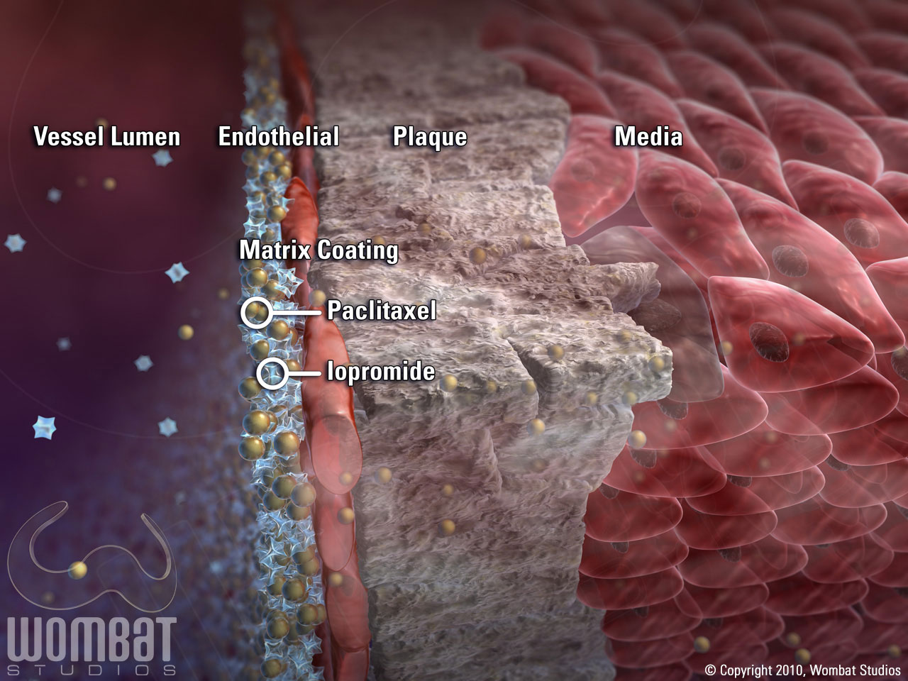
Paccocath: Close Up Image 1
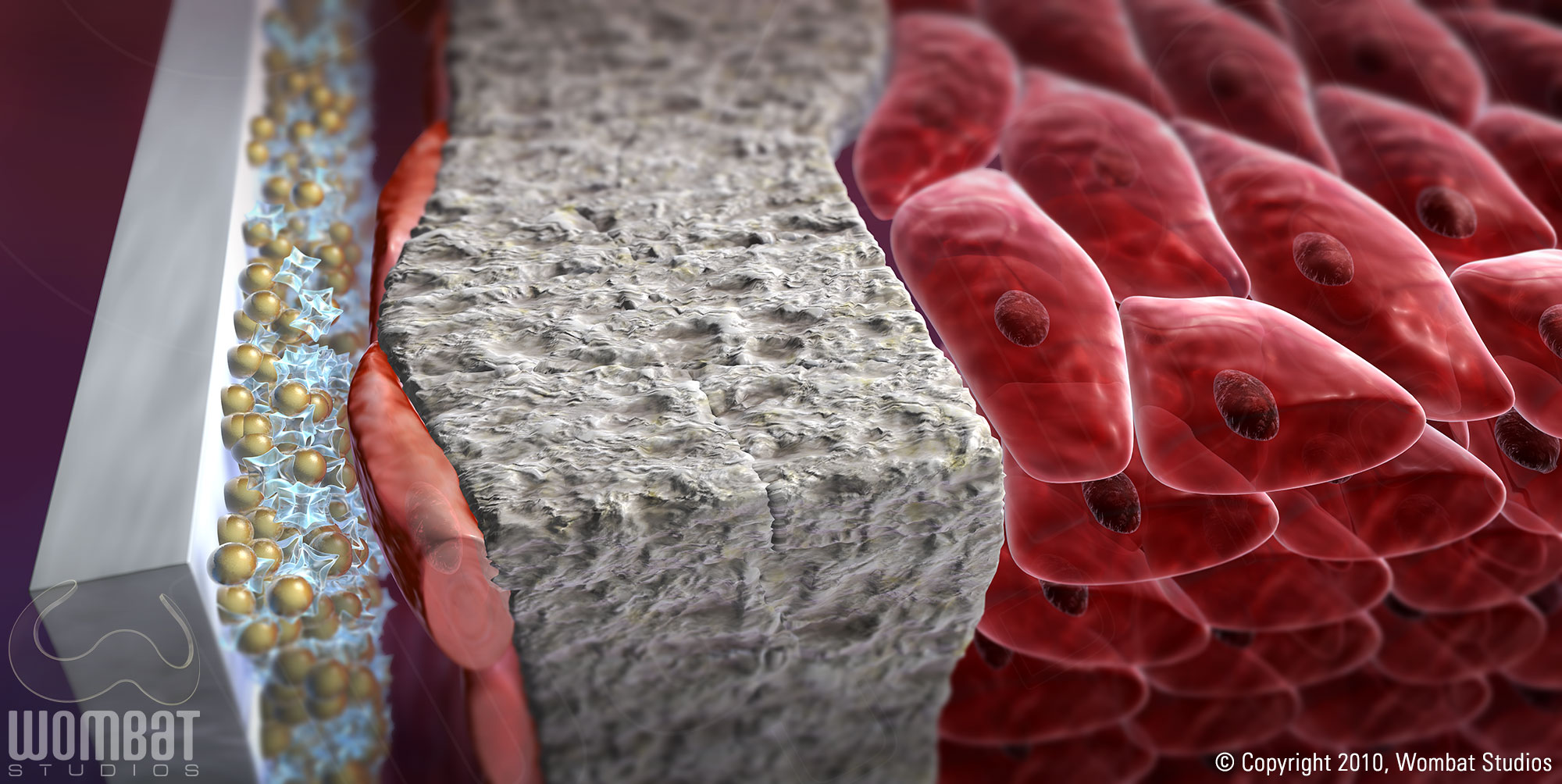
This is an up close, cross section view of the balloon, the drug coating, calcified plaque within the intima and medial cells of the vessel wall (the animation indicates these different elements through callouts). What we see here is the balloon just before it compresses the diseased vessel wall.
Paccocath: Close Up Image 2
When the balloon expands, the calcified plaque fractures and has the potential to damage or destroy cells, which in turn can cause uncontrolled regrowth, also known as restenosis (the animation shows this happening). The Paccocath drug coating is there to prevent restenosis. Here we see that the balloon has deflated, and much of the drug coating has remained on the lumen wall; there is some wash away (the particles we see floating to the left), but a significant portion of the drug is absorbed.
Paccocath: Drug Matrix
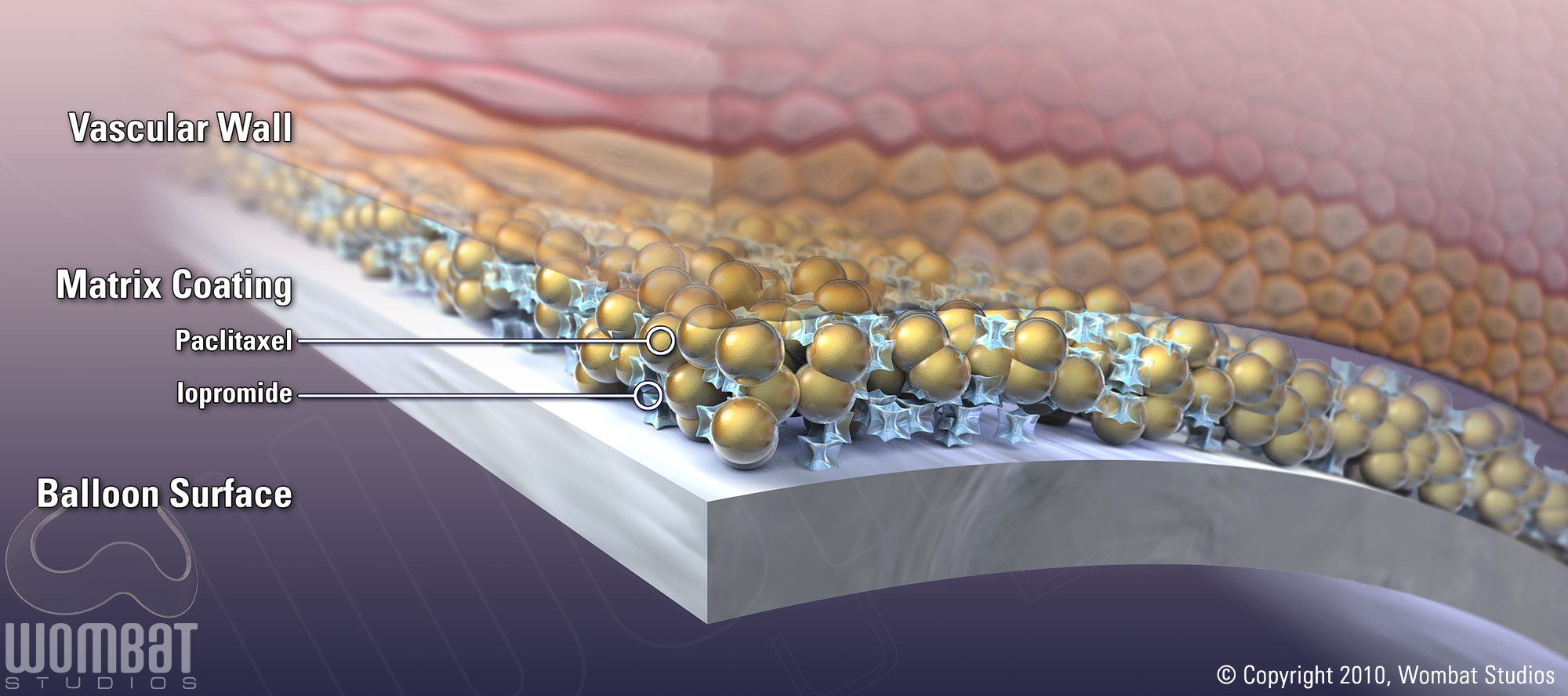
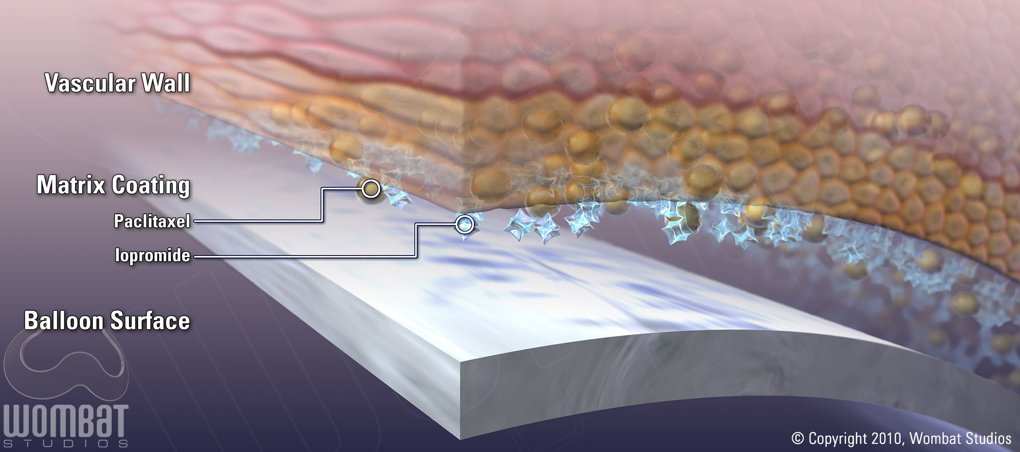
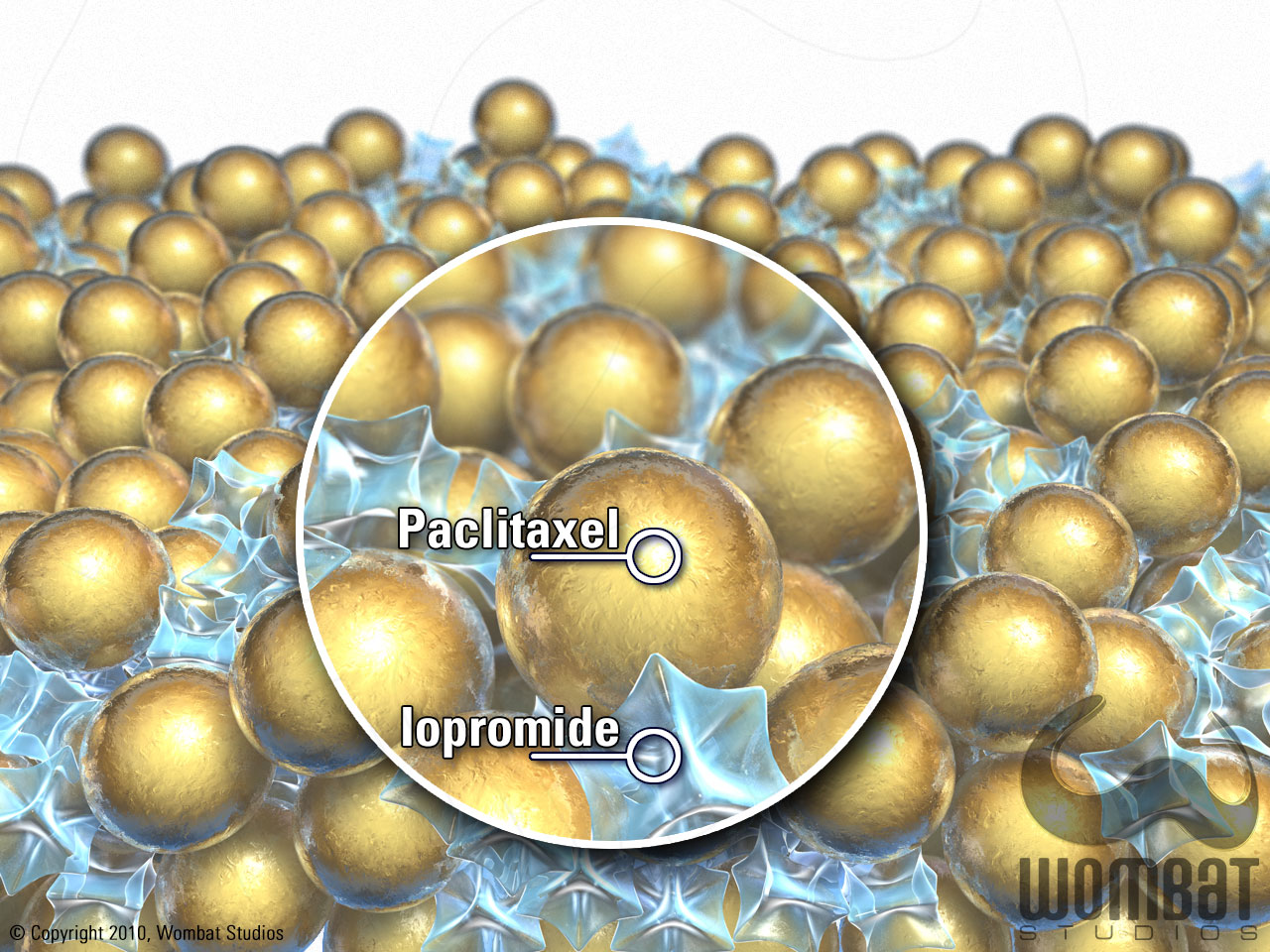
These images were created to feature the unique drug coating matrix, made up of Paclitaxel and Iopromide; they were originally created for presentation needs, but fit well into the animation, too.